Estudio microscópico del Bulbus oculi en cabras de Alepo (Damasco)
Resumen
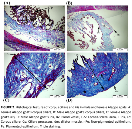
El ojo es uno de los órganos sensoriales más complejos y esenciales, responsable de percibir estímulos del entorno. Este estudio tuvo como objetivo examinar histológicamente la estructura ocular de la cabra (Capra hiscus) de Alepo (Damasco), una raza ampliamente criada en las regiones del Mediterráneo Oriental y el Sureste de Anatolia para la producción de leche y carne. Se recolectaron veinte bulbos oculares (bulbus oculi) de 10 cabras adultas (5 machos y 5 hembras) y se procesaron utilizando métodos histológicos estándar. El examen reveló la presencia de las tres túnicas principales del ojo, la túnica fibrosa (esclerótica y córnea), la túnica vascular (coroides, cuerpo ciliar e iris) y la túnica nerviosa (retina). La esclerótica estaba compuesta por tres capas, epiesclerótica, substantia propria y lámina fusca, con abundantes fibras de colágeno y melanocitos. La córnea se caracterizaba por un epitelio escamoso estratificado no queratinizado, la capa de Bowman, un estroma colagenoso, la membrana de Descemet y un endotelio. Notablemente, en la unión esclerocornea se identificaron el canal de Schlemm y la malla trabecular. La coroides contenía cinco capas distintas, incluyendo un tapetum fibrosum compuesto por haces de colágeno organizados pero sin melanocitos. El cuerpo ciliar y el iris mostraron un epitelio bicapa y estructuras musculares lisas bien desarrolladas. La retina estaba compuesta por diez capas, consistentes con las de otras especies de mamíferos, y no se observaron diferencias histológicas significativas entre cabras machos y hembras. Este estudio proporciona una referencia histológica fundamental sobre el ojo de la cabra de Alepo y puede contribuir a futuros estudios clínicos y experimentales relacionados con la salud ocular y la anatomía comparada.
Descargas
Citas
Eurell JA, Frappier BL. Dellmann’s textbook of Veterinary histology. 6th. ed. Ames, USA: Blackwell Publishing; 2006.
Akın F, Samsar E. Göz hastalıkları. 2nd. Ed. Ankara: Medipres; 2005.
Soliman SM, Adam ZAA, Abd Allah UKM. Light and electron microscopic structure of goat’s retina. J. Vet. Med. Res. [Internet]. 2010; 20(1):52-62. doi: https://doi.org/p8mp DOI: https://doi.org/10.21608/jvmr.2020.77580
Junqueira LC, Carneiro J. Basic histology. 10th. ed. New York: McGraw-Hill; 2003.
Nilsson DE. Eye evolution and its functional basis. Vis. Neurosci. [Internet]. 2013; 30(1-2):5-20. doi: https://doi.org/f24mdf DOI: https://doi.org/10.1017/S0952523813000035
Mavrogenis AP, Constantinou A, Louca A. Environmental and genetic causes of variation in production traits of Damascus goats. 1. Pre-weaning and post-weaning growth. Anim. Sci. [Internet]. 1984; 38(1):91-97. doi: https://doi.org/bzmjd8 DOI: https://doi.org/10.1017/S0003356100041398
Economides S, Georghiades E, Koumas A, Hadjipanayiotou M. The effect of cereal processing on the lactation performance of Chios sheep and Damascus goats and the pre-weaning growth of their offspring. Anim. Feed Sci. Technol. [Internet]. 1989; 26(1-2):93-104. doi: https://doi.org/crjktn DOI: https://doi.org/10.1016/0377-8401(89)90009-6
Constantinou A. Genetic and environmental relationships of body weight, milk yield and litter size in Damascus goats. Small Rumin. Res. [Internet]. 1989; 2(2):163-174. doi: https://doi.org/dsv4dn DOI: https://doi.org/10.1016/0921-4488(89)90041-2
Yalçın BC. Sheep and goats in Turkey. Rome: FAO; 1986 [cited 22 May 2025]. Available in: https://goo.su/UdfNUVf
Keskin M. Hatay Bölgesinde Yoğun Yetiştirme Koşullarında Şam (Damascus) Keçilerinin Morfolojik Özellikleri ve Performanslarının Saptanması [Doctoral thesis on the Internet]. Hatay: Mustafa Kemal Üniversitesi. 2000 [cited 22 May 2025] 103 p. Available in: https://goo.su/alRz8sT
Ohuchi H, Sato K, Habuta M, Fujita H, Bando T. Congenital eye anomalies: more mosaic than thought?. Congenital Anomalies. [Internet]. 2019; 59(3):56-73. doi: https://doi.org/p8mq DOI: https://doi.org/10.1111/cga.12304
Kirbaş-Doğan G, Koral-Taşçı S, Dalga S, İlhan-Aksu S. Anatomical and histological studies on the eyes of brown bear (Ursus arctos horribilis). Turk. J. Vet. Anim. Sci. [Internet]. 2020; 44(4):871–878. doi: https://doi.org/p8mr DOI: https://doi.org/10.3906/vet-2002-22
Okşar D, Orhan İ, Alan A, Köse F, Düzler A. Anatomical study of bulbus oculi in Akkaraman sheep. Erciyes Üniv. Vet. Fak. Derg. [Internet]. 2021; 18(3):145–151. doi: https://doi.org/p8ms DOI: https://doi.org/10.32707/ercivet.1015778
Jabbar AI, Ibrahim RS. Dıssector And Morphometrıc Study Of The Buffaloes Eyeballs (Bubalus Bubulıs). Biochem. Cell. Archiv. [Internet]. 2021 [cited Apr 23 2025]; 21(2):4303-4308. Available in: https://goo.su/KyiT
Dalga S, Aksu SI, Aslan K, Deprem T, Uğran R. Anatomical and histological structures of eye and lacrimal gland in Norduz andMorkaraman sheep. Turk. J. Vet. Anim. Sci. [Internet]. 2022; 46(2):336-346. doi: https://doi.org/p8mt DOI: https://doi.org/10.55730/1300-0128.4181
Dalga S, Aslan K, Taşçı SK, Ermutlu D, Aksu Sİ. Morphometric and Histological Structure of Bulbus Oculi in Goats. Acta Morphol. Anthropol. [Internet]. 2024; 31(1-2):94-108. doi: https://doi.org/p8mw DOI: https://doi.org/10.7546/AMA.31.1-2.2024.09
Yılmaz C, Kabak M, Selviler-Sizer S. Comparative macroanatomical and scanning electron microscopy study of the eyeball in brachycephalic and mesocephalic dog breeds. Microsc. Res. Tech. [Internet]. 2024; 87(10):2408-2417. doi: https://doi.org/p8mx DOI: https://doi.org/10.1002/jemt.24624
Öktay M. Comparative anatomy of vertebrate animals. İstanbul: İstanbul Üniversitesi Fen Fakültesi. [Internet]. 1998; p. 340–53. Turkish.
Ross N. Anatomy of domestic birds. Ankara: Medisan Publishing House; 2008.
Nickel R, Schummer A, Seiferle E, Wight PAL. The Anatomy of the Domestic Animals. Berlin: Springer; 2004.
Klećkowska-Nawrot JE, Goździewska-Harłajczuk K, Darska M, Barszcz K, Janeczek M. Microstructure of the eye tunics, eyelids and ocular glands of the Sulawesi bear cuscus (Ailurops ursinus Temminck, 1824) (Phalangeridae: Marsupialia) based on anatomical, histological and histochemical studies. Acta Zool. [Internet]. 2019; 100(2):182-210. doi: https://doi.org/gmsqnb DOI: https://doi.org/10.1111/azo.12251
Prince JH, Diesem CD, Eglitis I, Ruskall GL. Anatomy and histology of the eye and orbit in domestic animals. Springfield (IL): Charles C. Thomas; 1960.
König HE, Liebich HG. Anatomia zwierzat domowych, Kolorowy Atlas i Podrecznik. 2nd. ed. Łódź, Poland: Wydawnictwo Galaktyka; 2006.
Cafaro TA, Suarez MF, Maldonado C, Croxatto JO, Insfran C, Urrets-Zavalía JA, Serra HM. On the cornea of healthy merino sheep: a detailed ex vivo confocal, histological and ultrastructural study. Anat. Histol. Embryol. [Internet]. 2015; 44(4):247-254. doi: https://doi.org/f7km97 DOI: https://doi.org/10.1111/ahe.12131
Nautscher N, Bauer A, Steffl M, Amselgruber WM. Comparative morphological evaluation of domestic animal cornea. Vet. Ophthalmol. [Internet]. 2016; 19(4):297-304. doi: https://doi.org/f86sbs DOI: https://doi.org/10.1111/vop.12298
Merindano MD, Costa J, Canals M, Potau JM, Ruano D. A comparative study of Bowman’s layer in some mammals: Relationships with other constituent corneal structures. Eur. J. Anat. [Internet]. 2002 [cited Jun 15 2025]; 6(3):133-139. Available in: https://goo.su/JGFtV
Rehbinder C, Winquist G, Roos C. Structure of the cornea in some cervidae. Acta Vet. Scand. [Internet]. 1977; 18(2):152-158. doi: https://doi.org/p8m3 DOI: https://doi.org/10.1186/BF03548443
Reichard M, Hovakimyan M, Wree A, Meyer-Lindenberg A, Nolte I, Junghans C, Guthoff R, Stachs O. Comparative in vivo confocal microscopical study of the cornea anatomy of different laboratory animals. Curr. Eye Res. [Internet]. 2010; 35(12):1072-1080. doi: https://doi.org/c6cm8p DOI: https://doi.org/10.3109/02713683.2010.513796
Paszta W, Klećkowska-Nawrot JE, Goździewska-Harłajczuk K. Anatomical and morphometric evaluation of the orbit, eye tunics, eyelids and orbital glands of the captive females of the South African painted dog (Lycaon pictus pictus Temminck, 1820) (Caniformia: Canidae). PLoS one. [Internet]. 2021;16(4):e0249368. doi: https://doi.org/p8m5 DOI: https://doi.org/10.1371/journal.pone.0249368
Bacha Jr WJ, Bacha LM. Color Atlas of Veterinary Histology. 3rd. ed. Iowa, USA: John Wiley-Blackwell; 2012.
Slatter DH. Fundamento de Oftalmología Veterinaria. 3rd. ed. São Paulo: Roca; 2005.
Malsawmkima BR, Vyas YL, Bhayani DM. Histomorphological study on vascular tunics of the adult Surti buffalo (Bubalus bubalis). Int. J. Interdiscip. Multidiscip. Stud. [Internet]. 2014 [cited 22 May 2025]; 2:24-28. Available in: https://goo.su/R7tes
Schwab IR, Yuen CK, Buyukmihci NC, Blankenship TN, Fitzgerald PG. Evolution of the tapetum. Trans. Am. Ophthalmol. Soc. [Internet]. 2002 [cited 12 May 2025]; 100:187-200. Available in: https://goo.su/UhQg
Ollivier FJ, Samuelson DA, Brooks DE, Lewis PA, Kallberg ME, Komáromy AM. Comparative morphology of the tapetum lucidum (among selected species). Vet. Ophthalmol. [Internet]. 2004; 7(1):11-22. doi: https://doi.org/bkn4mc DOI: https://doi.org/10.1111/j.1463-5224.2004.00318.x
Aly KH, Imam HE. Comparative morphological studies on the vascular tunic of the eyeball in some of two species of fishes: Oreochromis Niloticus and Mugil Cephalus. [Thesis of Grade on the Internet]. Assiut: Assiut University. Faculty of Veterinary Medicine. Department of Anatomy and Histology; 2006 [cited 22 May 2025]. 12 p. Available in: https://goo.su/c4gk6
Derbalah AE. Light & Electron microscopical studies on the eye of one–humped camel (Camelus dromedaries). [Thesis of Grade on the Internet]. Alexandria: Alexandria University. Faculty of Veterinary Medicine. Department of Histology; 2001 [cited 22 May 2025]. 15 p. Available in: https://goo.su/OZRk4v
Ehrenhofer MCA, Deeg CA, Resse S, Liebich HG, Stangassinger M, Kaspers B. Normal structure and age- related changes of the equine retina. Vet. Ophthalmol. [Internet]. 2002; 5(1):39–47. doi: https://doi.org/bj3942 DOI: https://doi.org/10.1046/j.1463-5224.2002.00210.x
Altunay H. Fine structure of the retinal pigment epithelium, Bruch’s membrane and choriocapillaris in the ostrich (Struthio camelus). Anat. Histol. Embryol. [Internet]. 2004; 33:38–41. doi: https://doi.org/bjg5xm DOI: https://doi.org/10.1111/j.1439-0264.2004.00507.x
Samuelson DA. Textbook of Veterinary Histology. Philadelphia: Saunders (Elsevier); 2007.