Macroscopic and microscopic study of the brain structure of the common kestrel (Falco tinnunculus)
Abstract
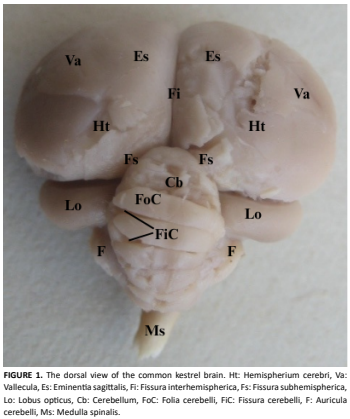
This study was conducted to examine the macroscopic and light microscopic structures of the brain in kestrels (Falco tinnunculus). In the study, brains from three adult kestrels that were brought in for treatment but could not be rescued, one from Keçiören Municipality Veterinary Affairs Directorate and two from Afyon Kocatepe University Wildlife Rescue and Rehabilitation Centerwere used. The brains were properly removed and subjected to macroscopic and light microscopic examination procedures. The average brain weight was measured as 3.23 g, total length as 22.5 mm, and total width as 23.5 mm. The cerebrum, cerebellum, lobus opticus, and medulla oblongata were the prominent regions of the brain. A well-developed lobus opticus was observed, and small and rudimentary bulbus olfactorius structures were present on both sides of the midline at the rostroventral part of the cerebral hemispheres. In the cerebellum, transverse protrusions called folia cerebelli, varying in number between 9 and 10, and the fissura cerebelli separating them were seen. On the sides of the cerebellum and behind the lobus opticus, auricula cerebelli were identified. At the caudal part of the brain, the pons and medulla oblongata were present and separated by a slightly prominent transverse groove. In the light microscopic examination, small neurons located beneath the piamater in the cerebral cortex and irregularly distributed glial cells among them were observed. In the deeper cortical regions, large neurons with vesicular nuclei were identified. In the cerebellum, a typical three-layered structure consisting of molecular, Purkinjecell, and granular layers was present. In addition, numerous multipolar neurons and supporting glial cells were found in the medulla oblongata, and ependymal cells were identified in the fourth ventricle. In the optic lobe, medium-sized spherical neurons containing few glial cells attracted attention. With this study, the macroscopic and light microscopic structures of the brain in kestrels were revealed. The data obtained were compared with the morphological structures of the brains of other avian species, and similarities and differences were discussed.The findings are expected to contribute to the literature on the morphological characteristics of raptors and serve as a foundation for comparative anatomical and histological studies on wild bird species.
Downloads
References
Nelson T. Falco tinnunculus (common kestrel). [Internet]. Animal Diversity Web: Tanya Dewey; 3 Feb 2006 [cited 3 Jul 2025]. Available in: https://goo.su/Iz8Uhl
Çakır A, Bakıcı C, Batur B. Evcil Kanatlı Hayvanların Anatomisi. 1st ed. Ankara: Ankara Nobel Tıp Kitabevleri; 2024.
Al-Nakeeb GD, Jasim NN. Morphological and Histological Study of the Forebrain (Cerebrum) in a Wild Bird Species (Columba livia domestica) (Gmelin, 1789). Baghdad Sci. J. [Internet]. 2018; 15(2):138-144. doi: https://doi.org/p92j DOI: https://doi.org/10.21123/bsj.15.2.138-144
Kardong KV. Vertebrates: Comparative Anatomy, Function, Evolution. 8th ed. New York: McGraw Hill; 2019.
Lőw P, Molnár K, Kriska G. Atlas of Animal Anatomy and Histology. 1st ed. Switzerland: Springer; 2016. doi: https://doi.org/p92k DOI: https://doi.org/10.1007/978-3-319-25172-1
Cobb S. Observations on the Comparative Anatomy of the Avian Brain. Perspect. Biol. Med. [Internet]. 1960; 3(3):383-408. doi: https://doi.org/p92m DOI: https://doi.org/10.1353/pbm.1960.0053
Abd-Alrahman SA. Morphological and Histological Study Of The Cerebrum In A Nocturnal Bird Species (Barn owl) Tyto Alba. Ibn AL-Haitham J. Pure. Appl. Sci. [Internet]. 2017 [cited 8 Jun 2025]; 25(3):73-87. Available in: https://goo.su/s8tv9R
Mohammed EAM, Jala HAN, Abd-Alhafid YKA. Anatomical study of the Cerebrum of the laughing dove (Stigmatopelia senegalensis). Glob. Libyan J. [Internet]. 2018 [cited 8 Jun 2025]; 39:1-9. Available in: https://goo.su/vzcZoV
Abid AB, Al-Bakri NA. Morphological and Histological Study of the Fore Brain (Cerebrum) In Quail Coturnix coturnix (Linnaeus, 1758). Ibn AL-Haitham J. Pure. Appl. Sci. [Internet]. 2016 [cited 1 Jul 2025]; 29(1):25-39. Available in: https://goo.su/9kIIUh
Azmat U, Nisar H, Shah SMR, Aziz H, Baig M, Irshad A, Rehmat, Sikandar M. Histo-Morphometrical Study of the Central Nervous System of Rose-Ringed Parakeet (Psittacula krameria) In Breeding and Non-Breeding Seasons. Saudi J. Pathol. Microbiol. [Internet]. 2024; 9(11):237-248. doi: https://doi.org/p92q DOI: https://doi.org/10.36348/sjpm.2024.v09i11.002
Joshi SK, Udgata J, Sathapathy S, Sahu SK. Gross morphological studies on the brain of Kadaknath fowl in growing period. J. Entomol. Zool. Stud. [Internet]. 2019 [cited 1 Jul 2025]; 7(5):353-355. Available in: https://goo.su/cseKhi
Aslan Ş, Deprem T, Bingöl SA, Koral-Taşçı S. Kanatlı Histolojisi. 1st ed. Bursa: Dora Yayıncılık; 2018.
Elnegiry AA, Hamoda HS, Farrag FA. Histomorphological Study on the Cerebellum of the African Ostrich. Alex. J. Vet. Sci. [Internet]. 2022; 73(2):1-6. doi: https://doi.org/p92r DOI: https://doi.org/10.5455/ajvs.33484
Baumel JJ, King SA, Breasile JE, Evans HE, Berge JCV. Handbook of avian anatomy: nomina anatomica avium. 2nd ed. Cambridge MA, EEUU: Nuttall Ornithological Club;1993.
König HE, Korbel R, Liebich HG. Avian Anatomy: Textbook and Colour Atlas. 2nd ed. Sheffield, UK: 5m Publishing; 2016.
Balkaya H, Toprak B. External anatomical structures of sparrowhawk (Accipiter nisus) encephalon. Indian J. Anim. Res. [Internet]. 2018; 52(9):1281-1284. doi: https://doi.org/p92t
Batah AL, Ghaje MS, Sh. NA. Anatomical and Histological study for the Brain of the locally breed chicken (Gallus gallus domesticus). J. Thi-Qar Sci. [Internet]. 2012 [cited 8 Jun 2025]; 3(3):47-53. Available in: https://goo.su/4I8XbR
Peng KM, Feng Y, Zhang G, Liu H, Song H. Anatomical study of the brain of the African ostrich. Turk. J. Vet. Anim. Sci. [Internet]. 2010; 34(3):235-241. doi: https://doi.org/p92z
Taşbaş M. Evcil Kanatlılardan Tavuk-Horoz (Gallus domesticus) ve Hindi’nin (Meleagris gallopavo) Encephalon ve Zarları (Meninges) Üzerinde Karşılaştırmalı Makro-Anatomik Ve Subgros Araştırmalar. Ankara Univ. Vet. Fak. Derg. [Internet]. 1978; 25(4): 747-759. doi: https://doi.org/p922 DOI: https://doi.org/10.1501/Vetfak_0000001139
Karkoura A, Alsafy M, El-gendy S, El-defrawy F. Morphological Investigation of the Brain of the African Ostrich (Struthio camelus). Int. J. Morphol. [Internet]. 2015; 33(4):1468-1475. doi: https://doi.org/p923 DOI: https://doi.org/10.4067/S0717-95022015000400046
Hall ZJ, Street SE, Healy SD. The evolution of cerebellum structure correlates with nest complexity. Biol. Lett. [Internet]. 2013; 9(6):20130687. doi: https://doi.org/gb9fv2 DOI: https://doi.org/10.1098/rsbl.2013.0687
Koral-Taşçı S. Histological and Histometric Structure of Goose (Anser anser) Cerebellum. Van Vet. J. [Internet]. 2018 [cited 8 Jun 2025]; 29(2):63-66. Available in: https://goo.su/DixPrd
Sur E, Öznurlu Y, Özaydın T, Çolakoğlu F, Ünsal S, Yener Y. Comparative histometrical study of the cerebellum and the determination of some AgNOR parameters in different avian species. Bull. Vet. Inst. Pulawy. [Internet]. 2011 [cited 8 Jun 2025]; 55:261-265. Available in: https://goo.su/RT5r8kE
Vincze O, Vágási CI, Pap PL, Osváth G, Møller AP. Brain regions associated with visual cues are important for bird migration. Biol. Lett. [Internet]. 2015; 11(11):20150678. doi: https://doi.org/p924 DOI: https://doi.org/10.1098/rsbl.2015.0678
Gupta SK, Behera K, Pradhan CR, Mandal AK, Sethy K, Behera D, Shinde KP. Studies of the macroscopic and microscopic morphology (hippocampus) of brain in Vencobb broiler. Vet. World. [Internet]. 2016; 9(5):507-511. doi: https://doi.org/p925 DOI: https://doi.org/10.14202/vetworld.2016.507-511
Hussain RSH, Al-taee AA. Comparative Study between Brain and Optic Lobe of Falcon (Falco columbarius) and Owl (Bubo bubo). Pak. J. Med. Health Sci. [Internet]. 2022; 16(4):909-912. doi: https://doi.org/p926 DOI: https://doi.org/10.53350/pjmhs22164909
Bang BG, Cobb S. The Size of the Olfactory Bulb in 108 Species of Birds. The Auk. [Internet]. 1968; 85(1):55-61. doi: https://doi.org/p927 DOI: https://doi.org/10.2307/4083624
Abankwah V, Deeming D, Pike T. Avian olfaction: a review of the recent literature. Comp. Cogn. Behav. Rev. [Internet]. 2020; 15:149-161. doi: https://doi.org/p928 DOI: https://doi.org/10.3819/CCBR.2020.150005
Jones HC, Dolman GS. The structure of the roof of the fourth ventricle in pigeon and chick brains by light and electron microscopy. J. Anat. [Internet]. 1979 [cited 8 Jun 2025]; 128(Pt 1):13-29. Available in: https://goo.su/Arzr