Aplicación de Deep Learning para el diagnóstico rápido y preciso de fracturas óseas en perros mediante imágenes radiográficas
Resumen
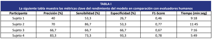
La radiografía continúa siendo la herramienta diagnóstica de mayor uso en la medicina veterinaria para la detección de fracturas óseas en perros. No obstante, su interpretación manual puede verse afectada por la experiencia del profesional, así como por factores como el cansancio o la sobrecarga laboral. En este contexto, el presente estudio evaluó el desempeño de un modelo de deep learning basado en la arquitectura YOLOv5, orientado al diagnóstico de imágenes radiográficas caninas divididas en dos categorías: presencia o ausencia de fracturas. El modelo alcanzó una precisión del 83.3%, superando significativamente a tres médicos veterinarios generales, quienes registraron porcentajes de acierto de entre el 40% y el 70%. Además, el sistema automatizado permitió reducir el tiempo promedio de diagnóstico en un 40%, logrando clasificaciones en cuestión de segundos. Esos resultados resaltan la viabilidad de la inteligencia artificial como herramienta para mejorar la precisión, la rapidez y la consistencia del diagnóstico en medicina veterinaria, especialmente en entornos con recursos humanos limitados. Se recomienda ampliar la base de datos y validar el modelo en contextos clínicos reales.
Descargas
Citas
Couch JR. Artificial Intelligence: Past, Present and Future. J. SC Acad. Sci. [Internet]. 2023 [citado 08 Enero 2025]; 21(1):2. Disponible en: https://goo.su/SYEdM
Anwar SM, Majid M, Qayyum A, Awais M, Alnowami M, Khan MK. Medical Image Analysis using Convolutional Neural Networks: A Review. J. Med. Syst. [Internet]. 2018; 42(11):226. doi: https://doi.org/gfghhd DOI: https://doi.org/10.1007/s10916-018-1088-1
Appleby RB, Basran PS. Artificial intelligence in veterinary medicine. J. Am. Vet. Med. Assoc. [Internet]. 2022; 260(8):819–824. doi: https://doi.org/p7f6 DOI: https://doi.org/10.2460/javma.22.03.0093
Davenport T, Kalakota R. The potential for artificial intelligence in healthcare. Future Healthc. J. 2019; 6(2):94–98. doi: https://doi.org/ggf26q DOI: https://doi.org/10.7861/futurehosp.6-2-94
Rajpurkar P, Irvin J, Zhu K, Yang B, Mehta H, Duan T, Ding D, Bagul A, Langlotz C, Shpanskaya K, Lungren MP, Ng AY. CheXNet: Radiologist-Level Pneumonia Detection on Chest X-Rays with Deep Learning. Cornell University: Arxiv; 2018 [citado 08 Enero 2025]. doi: https://doi.org/g88dtm
Lubinus Badillo F, Rueda Hernández CA, Marconi Narváez B, Arias Trillos YE. Redes neuronales convolucionales: un modelo de Deep Learning en imágenes diagnósticas. Revisión de tema. Rev. Colomb. Radiol. 2021; 32(3):5591– 5599. doi: https://doi.org/p7f7 DOI: https://doi.org/10.53903/01212095.161
Mora-Tola MA, Carpio-Alemán FM, Mora-Tola JD, Román-Cárdenas FA. Characterization of fractures of the appendicular skeleton in dogs according to the AO classification. Polo Conoc. 2023 [citado 08 Enero 2025]; 8(3):2440–2457. Disponible en: https://goo.su/4fgGIK6
DeCamp CE, Johnston SA, Dejardin LM, Schaefer SL. Brinker, Piermattei and Flo’s Handbook of Small Animal Orthopedics and Fracture Repair. 5th ed. St. Louis: Saunders; 2016.
McEvoy FJ, Amigo JM. Using machine learning to classify image features from canine pelvic radiographs: evaluation of partial least squares discriminant analysis and artificial neural network models. Vet. Radiol. Ultrasound. 2013; 54(2):122–126. doi: https://doi.org/f4rr93 DOI: https://doi.org/10.1111/vru.12003
Shen D, Wu G, Suk HI. Deep learning in medical image analysis. Annu. Rev. Biomed. Eng. 2017; 19(1):221–248. doi: https://doi.org/gcgmb4 DOI: https://doi.org/10.1146/annurev-bioeng-071516-044442
Rajpurkar P, Irvin J, Ball RL, Zhu K, Yang B, Mehta H, Duan T, Ding D, Bagul A, Langlotz CP, Patel BN, Yeom KW, Shpanskaya K, Blankenberg FG, Seekins J, Amrhein TJ, Mong DA, Halabi SS, Zucker EJ, Ng AY, Lungren MP. Deep learning for chest radiograph diagnosis: a retrospective comparison of the CheXNeXt algorithm to practicing radiologists. PLoS Med. 2018; 15(11):e1002686. doi: https://doi.org/gfnkcv DOI: https://doi.org/10.1371/journal.pmed.1002686
Taha AA, Hanbury A. Metrics for evaluating 3D medical image segmentation: analysis, selection, and tool. BMC Med. Imaging. 2015; 15(1):29. doi: https://doi.org/gb343z DOI: https://doi.org/10.1186/s12880-015-0068-x
Lakhani P, Sundaram B. Deep learning at chest radiography: automated classification of pulmonary tuberculosis by using convolutional neural networks. Radiology. 2017; 284(2):574–582. doi: https://doi.org/gbp274 DOI: https://doi.org/10.1148/radiol.2017162326
Schmidhuber J. Deep learning in neural networks: an overview. Neural Netw. 2015; 61:85–117. doi: https://doi.org/f6v78n DOI: https://doi.org/10.1016/j.neunet.2014.09.003
Nature Machine Intelligence. A prize for discoveries past, present and future. Nat. Mach. Intell. [Internet]. 2019; 1(5):201. doi: https://doi.org/p7hf DOI: https://doi.org/10.1038/s42256-019-0054-z
Netter A, Noorzadeh S, Duchateau F, Abrao H, Desternes J, Peyras J, et al. Initial Results in the Automatic Visual Recognition of Endometriosis Lesions by Artificial Intelligence During Laparoscopy: A Proof-of-Concept Study. J Minim Invasive Gynecol. 2025 Sep 3. Online. doi: https://doi.org/10.1016/j.jmig.2025.08.027 DOI: https://doi.org/10.1016/j.jmig.2025.08.027
Kukartsev VV, Ageev RA, Borodulin AS, Gantimurov AP, Kleshko II. Deep learning for object detection in images: development and evaluation of the YOLOv8 model using Ultralytics and Roboflow libraries. In: Silhavy R, Silhavy P, editors. Software Engineering Methods Design and Application. CSOC 2024. Lecture Notes in Networks and Systems. Cham: Springer; 2024. p. 630. doi: https://doi.org/p7hg DOI: https://doi.org/10.1007/978-3-031-70285-3_48
Gulshan V, Peng L, Coram M, Stumpe MC, Wu D, Narayanaswamy A, Venugopalan S, Widner K, Madams T, Cuadros J, Kim R, Raman R, C. Nelson P, Mega JL, Webster DR. Development and validation of a deep learning algorithm for detection of diabetic retinopathy in retinal fundus photographs. JAMA. 2016; 316(22):2402-2410. doi: https://doi.org/gcgk7d DOI: https://doi.org/10.1001/jama.2016.17216
Guarnido-Lopez P, Ramirez-Agudelo JF, Denimal E, Benaouda M. Programming and setting up the object detection algorithm YOLO to determine feeding activities of beef cattle: A comparison between YOLOv8m and YOLOv10m. Animals. 2024; 14(19):2821. doi: https://doi.org/p7hh DOI: https://doi.org/10.3390/ani14192821
Abeliuk A, Gutiérrez C. Historia y evolución de la inteligencia artificial. Rev. Bits Cienc. 2021; 21:14-21. doi: https://doi.org/p7hj
Bengio Y, LeCun Y, Hinton G. Deep learning for AI. Commun ACM. 2021; 64(7):58–65. doi: https://doi.org/gkx7tb DOI: https://doi.org/10.1145/3448250
Esteva A, Kuprel B, Novoa RA, Ko J, Swetter SM, Blau HM, Thrun S. Dermatologist-level classification of skin cancer with deep neural networks. Nature. 2017; 542(7639):115–118. doi: https://doi.org/bxwn DOI: https://doi.org/10.1038/nature21056
Gerke S, Minssen T, Cohen G. Ethical and legal challenges of artificial intelligence-driven healthcare. In: Bohr A, Memarzadeh K, editors. Artificial Intelligence in Healthcare. Amsterdam: Academic press; 2020. p. 295–336. doi: https://doi.org/gxww DOI: https://doi.org/10.1016/B978-0-12-818438-7.00012-5
He K, Zhang X, Ren S, Sun J. Deep residual learning for image recognition. In: Proceedings of the IEEE Conference on Computer Vision and Pattern Recognition (CVPR); Las Vegas, NV, USA; 2016. p. 770–778. doi: https://doi.org/gdcfkn DOI: https://doi.org/10.1109/CVPR.2016.90
Evans HE, de Lahunta A. Miller’s Anatomy of the Dog. 4th ed. St. Louis: Saunders. Can. Vet. J. 2012; 57(4):381. Available in: https://goo.su/yiOz0
Irvin J, Rajpurkar P, Ko M, Yu Y, Ciurea-Ilcus S, Chute C, Marklund H, Haghgoo B, Ball R, Shpanskaya K, Seekins J, Mong DA, Halabi SS, Sandberg JK, Jones R, Larson DB, Langlotz CP, Patel BN, Lungren MP, Ng AY. CheXpert: A large chest radiograph dataset with uncertainty labels and expert comparison. Proc. AAAI. Conf. Artif. Intell. 2019; 33(01):590–597. doi: https://doi.org/ghkh8x DOI: https://doi.org/10.1609/aaai.v33i01.3301590