Macroscopic and microscopic structure of the infraorbital sinus gland in Tuj Sheep
Abstract
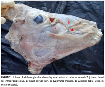
The infraorbital sinus (infraorbital gland-preorbital gland) is located in the lateral aspect of the skull, rostroventral to the eye, within the infraorbital fossa. Some skin glands are differentiated and specialized. In the present study, it was aimed to examine the macroscopic and microscopic structure of the infraorbital sinus, which is one of these skin glands. The distance between the infraorbital foramen and infraorbital sinus was 47.35± 0.57mm on the right and 53.80±1.27 mm on the left. The left side length of the infraorbital sinus in Tuj sheep was measured as 25.32±2.21 mm, width 16.21±2.83 mm, and depth 1.23±1.80 mm. The Tuj sheep infraorbital sinus was composed of the epidermis and dermis. The epidermis was consist of keratinized stratified squamous epithelium. There were many sebaceous glands, some apocrine glands, muscle fibers, and also hair follicles in the dermis. The sebaceous glands were located in the superficial region and the apocrine glands were deeper in the dermis. It was seen lots of lipid droplets inside the sebaceous gland cells. The myoepithelial cells and the basal membrane of the apocrine glands and muscle fibers were PAS-positive reaction. In conclusion, in the presented study, the macroscopic and microscopic structure of the infraorbital sinus was determined in Tuj sheep. This region is also clinically important because it is close to the infraorbital nerve passing through the infraorbital foramen (anesthesia area) and the infraorbital sinus is topographically located in this region. It is thought that it will support scientific studies related to the skin and its appendages.
Downloads
References
Demiraslan Y, Orhun-Dayan M. Veteriner Sistematik Anatomi. 1st. ed. Ankara, Turkey: Atlas Kitabevi; 2021.
Demiraslan Y, Orhun-Dayan M. VETKA-Veteriner Klinik Anatomi. 1st. ed.Ankara, Turkey: Nobel Akademik Yayincilik; 2023.
König HE, Liebich HG. Veteriner Anatomi (Evcil Memeli Hayvanlar). 3rd. ed.Turkey: Medipres; 2022.
Haziroglu RM, Cakir A, editors. Veteriner Anatomi Konu Anlatımı ve Atlas. 4th. ed. Ankara: Güneş Tip Kitabevleri; 2017.
Moawad UK. Morphological, histochemical and morphometric studies of the preorbital gland of adult male and female egyptian native breeds of sheep (Ovis aries).Asian J. Anim. Vet. Adv. [Internet]. 2016; 11(12):771-782. doi: https://doi.org/p73r. DOI: https://doi.org/10.3923/ajava.2016.771.782
King N, Kumar AHS, Kilroy D. Ovine infraorbital (preorbital) pouch: an unusual presentation in a male lamb.Biol. Eng. Med. Sci. Rep. [Internet]. 2018; 4(1):01-02. doi: https://doi.org/p73s. DOI: https://doi.org/10.5530/bems.4.1.1
Rajagopal T, Archunan G. Histomorphology of preorbital gland in territorial and non-territorial male blackbuck Antelope cervicapra, a critically endangered species.Biol. [Internet]. 2011; 66:370-378. doi: https://doi.org/cj22wj. DOI: https://doi.org/10.2478/s11756-011-0015-4
Dursun N. Veteriner Anatomi II. 12th. ed. Ankara: MedisanPublishing House; 2008.
Delmann HD, Brown EM. Textbook of Veterinary Histology. 2nd. Ed. Philapelphia: Lea & Febiger; 1981.
Atoji Y, Yamamoto Y, Suzuki Y. Infraorbital glands of a male formosan serow (Capricornis crispus swinhoei). Eur. J. Morphol. [Internet]. 1996; 34(2):87-94. doi: https://doi.org/fpczwb. DOI: https://doi.org/10.1076/ejom.34.2.87.13015
Ceaceroa F, Pluhácek J, Komárková M, Zábranský M. Pre- orbital gland opening during aggressive interactions in Rusa deer (Rusa timorensis). Behav. Proces. [Internet]. 2015; 111:51–54. doi: https://doi.org/f62ppw. DOI: https://doi.org/10.1016/j.beproc.2014.11.017
N.A.V. International Committee on Veterinary Gross Anatomical Nomenclature. Nomina Anatomica Veterinaria (I.C.V.G.A.N.). 6th. ed. World Association of Veterinary Anatomists: Hanover (Germany), Ghent (Belgium), Columbia, MO (U.S.A.), Rio de Janeiro (Brazil); 2017.
Liu M, Yang Y, Li M, Cai Y, Zhao S, Wang X. Comparison study of morphology andvascular casts between plateau- type Tibetan sheep andlow-altitude small-tailed Han sheep testes. Anat. Histol. Embryol. [Internet]. 2022; 51(4):524–532. doi: https://doi.org/p74r. DOI: https://doi.org/10.1111/ahe.12822
Adnyane IKM, Zuki ABZ, Noordin MM, Wahyuni S, Agungpriyono S. Morphological study of the infraorbital gland of the male barking deer, muntiacus muntjak. Afr. J. Biotechnol. [Internet]. 2011; 10(77):17891-17897. Available in: https://goo.su/TOR4k. DOI: https://doi.org/10.5897/AJB10.2634
Clutton-Brock T, Guinness FE, Albon SD. Red Deer, Behaviour and Ecology of Two Sexes. 1st. ed. Chicago, IL: University of Chicago Press; 1982.
Bartos L. Some observations on the relationship between preorbital gland opening and social interactions in red deer. Aggress. Behav. [Internet]. 1983; 9(1):59–67. doi: https://doi.org/dvpzk2. DOI: https://doi.org/10.1002/1098-2337(1983)9:1<59::AID-AB2480090108>3.0.CO;2-F
Hatlapa HH. Zur biologischen Bedeutung des praorbital Organs beim Rotwild, Pragung, Individualgeruch, Orientierung. Berl. Munch. Tierartzl. Wochenschr. 1977; 90:100–104.
Wolfel H. Zur Jugendentwicklung, Mutter-Kind-Bindung und Feindvermeidung beim Rothirsch (Cervus elaphus) II. Z. Jagdwissensch. [Internet]. 1983; 29:197–213. doi: https://doi.org/dgdq8t. DOI: https://doi.org/10.1007/BF02243645
Bartos L, Víchová J, Lancingerová J. Preorbital gland opening in red deer (Cervus elaphus) calves: Signals of hunger?. J. Anim. Sci. [Internet]. 2005; 83(1):124–129. doi: https://doi.org/p746. DOI: https://doi.org/10.2527/2005.831124x
Eurell JA, Frappier BL. Dellmann’s Textbook of Veterinary Histology. 6th. ed. Iowa (USA), Oxford (UK): Blackwell Publishing; 2006.
Gernek WH, Cohen M. The micromorphology of the glands of the infra-orbital cutaneous sinus of the Steenbok (Raphıcerus Campestrıs). Onderstepoort J. Vet. Res. [Internet]. 1978; 45(2):59-66.
Mohamed R, Driscoll M, Mootoo N. Clinical anatomy of the skull of the Barbados Black Belly sheep in Trinidad. Int. J. Curr. Res. Med. Sci. [Internet]. 2016; 2(8):8-19. Available in: https://goo.su/fQ9X.


